

1st Prize Winners – Dr. Jennifer Peters’ and Dr. Michael Taylor’s winning image of the blood-brain barrier in a live zebrafish embryo perfectly demonstrates the intersection of art and science that drives the Nikon Small World Competition.
The Nikon International Small World Competition first began in 1975 as a means to recognize and applaud the efforts of those involved with photography through the light microscope. Since then, Small World has become a leading showcase for photomicrographers from the widest array of scientific disciplines.
A photomicrograph is a technical document that can be of great significance to science or industry. But a good photomicrograph is also an image whose structure, color, composition, and content is an object of beauty, open to several levels of comprehension and appreciation. Nikon’s Small World is regarded as the leading forum for showcasing the beauty and complexity of life as seen through the light microscope. The Photomicrography Competition is open to anyone with an interest in microscopy and photography. Nikon recently announced the winners of the ‘2012 Photomicrography Competition’ judged by a panel of eminent professors and editors. Below are some of the photographs from the winning entries
 2nd Prize Winner -Walter Piorkowski for Live newborn lynx spiderlings (6x)
 3rd Prize Winner -Dr. Dylan Burnette for Human bone cancer (osteosarcoma) showing actin filaments (purple), mitochondria (yellow), and DNA (blue) (63x)
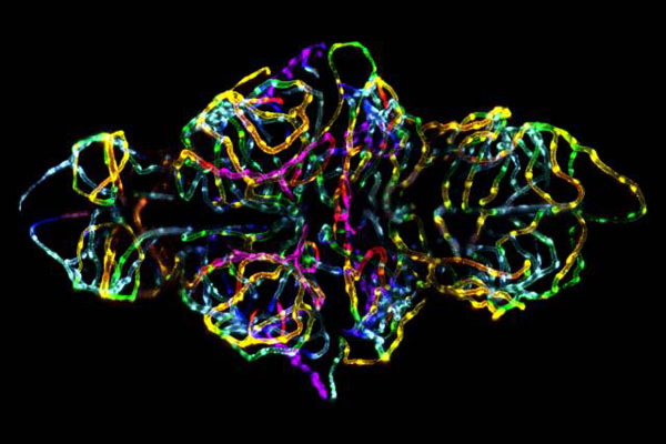
 4th Prize Winner -Dr. W. Ryan Williamson for Drosophila melanogaster visual system halfway through pupal development, showing retina (gold), photoreceptor axons (blue), and brain (green) (1500x)
 5th Prize Winner -Honorio Cócera-La Parra for Cacoxenite (mineral) from La Paloma Mine, Spain (18x)
 7th Prize Winner -Dr. Michael John Bridge for Eye organ of a Drosophila melanogaster (fruit fly) third-instar larvae (60x)
 12th Prize Winner – Esra Guc for 3D lymphangiogenesis assay. Cells sprout from dextran beads embedded in fibrin gel. (200x)
If you wish to participate in the 2013 Photomicrography competition, you can upload your works at MicroscopyU.com before 30th April 2013. for further details, visit Small World’s 2013 Photomicrography competition